Stroke
Compassionate and Rehabilitative Treatments Following a Stroke
Jupiter Medical Center treats hundreds of stroke patients annually. When
it comes to stroke, our team knows that "time is brain." We
offer a higher level of stroke expertise, as an accredited program, we
stay on the leading edge of stroke diagnosis and treatment. Because of
this level of expertise, more and more people are surviving and thriving
after acute stroke.
At Jupiter Medical Center, our state-of-the-art Stroke Center is equipped
with sophisticated technology to diagnose, intervene, and treat strokes
quickly. Our world-class center has everything you need in one location,
available around the clock.
If you think you or a loved one is suffering a stroke, call 911. If you
would like education on stroke risks or symptoms, contact our Stroke Center
today. We can be reached at
561-263-5972, or you can
find a physician here.
Types of Stoke, Warning Signs, and Risk Factors
The Ischemic Stroke is the most common type of stroke and occurs when an artery becomes blocked in the brain. Hemorrhagic strokes are the second most common type of stroke and occur as a response of a brain bleed that puts pressure on the brain cells, resulting in damage. Understanding risks and warning signs may help you better understand if you or a loved one is showing symptoms of having a stroke.
Stroke risk factors may include:
- Family history of strokes
- Age (55 and above increases risk)
- Previous history of stroke or ministroke
- Certain genetic conditions
- Smoking cigarettes
- High blood pressure
- Heart disease
- High cholesterol
- Diabetes
- Peripheral artery disease
- Untreated atrial fibrillation
- Obesity
Warning signs of a stroke may include:
- Sudden numbness or weakness of the face, arm, or leg, especially on one side of the body
- Sudden confusion, trouble speaking, or understanding
- Sudden trouble seeing in one or both eyes
- Sudden dizziness, trouble walking, or loss of balance/coordination
- Sudden severe headache with no known cause
You may have some or all of these signs. Be sure to note the time when symptoms start and call 911 immediately. Don’t ignore these warning signs, even if they go away. The symptoms of a stroke come on suddenly and should be emphasized as an emergency.
An easy way to remember how to recognize stroke symptoms is to B.E. F.A.S.T:
Balance – sudden loss of balance?
Eyes – loss of vision in one or both eyes?
Face drooping – face looks uneven?
Arm weakness – arm or leg weak, hanging down?
Speech – slurred speech? trouble speaking or seems confused?
Time – call 911 now!
There are treatments that can reduce the risk of damage from a stroke, but only if you get help quickly. Time is particularly critical because every second counts!
Jupiter Medical Center’s Stroke Center
As a Joint Commission designated Thrombectomy-Capable Stroke Center, we stay on the leading edge of stroke diagnosis and treatment, and our team is specially trained to manage the most complex conditions. This certification is a collaboration between The Joint Commission and the American Heart Association/American Stroke Association (AHA/ASA), which recognizes hospitals that have met rigorous standards for performing endovascular thrombectomy and providing post-procedural care.
 To become a Joint Commission certified Thrombectomy-Capable Stroke Center,
a program must meet ten core measures. These evidence-based measures are
considered best practice and were developed under the guidance of the
American Heart Association/American Stroke Association. Jupiter Medical
Center’s program is 100% compliant with all measures, meaning our
Stroke Center meets rigorous standards for managing acute stroke emergencies.
This includes:
To become a Joint Commission certified Thrombectomy-Capable Stroke Center,
a program must meet ten core measures. These evidence-based measures are
considered best practice and were developed under the guidance of the
American Heart Association/American Stroke Association. Jupiter Medical
Center’s program is 100% compliant with all measures, meaning our
Stroke Center meets rigorous standards for managing acute stroke emergencies.
This includes:
- Advanced imaging, medical equipment, and facilities
- Neurological intensive care units (ICU) for critical care
- Neurosurgical operating rooms
- Step-down units or close monitoring as your condition improves
- Doctors and other specialists trained in vascular (blood vessel), neurology, neurosurgery, and endovascular (minimally invasive) treatments
- Experience and knowledge of how to treat patients with complex types of strokes
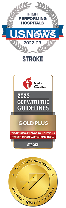
The American Heart Association (AHA/ASA) has also awarded our hospital with the Get with the Guidelines® – Stroke GOLD PLUS honor roll elite plus award, which represents the highest levels of achievement in key quality measures. Furthermore, the U.S. News & World Report, the trusted source in hospital rankings and consumer advice, has named Jupiter Medical Center a 2022-2023 High Performing hospital for Stroke. This is the highest award a hospital can earn for U.S. News' Best Hospitals Procedures & Conditions ratings. The annual Procedures & Conditions ratings are designed to assist patients and their doctors in making informed decisions about where to receive care for challenging health conditions or elective procedures.
Comprehensive Care When It Matters Most
A stroke can be overwhelming. There is a large amount of new information
to process, decisions to be made concerning treatment and care, and uncertainty
about which steps to take next or where to go for assistance. This often
leads to enormous stress for patients and their families.
At Jupiter Medical Center you don’t have to face these challenges
alone – we can help. Our
Stroke Coordinator can help you and your family – providing the information and guidance
needed to manage your stroke diagnosis and treatment. In addition to your
Stroke Coordinator, there is an entire team on your side to assist in
the treatment and recovery of stroke, for a truly multifaceted approach
to care. Our physicians are trained in neurology,
neuro-interventional radiology, and neurosurgery, and are qualified to provide advanced methods of treatment.
Our
state-of-the-art CT and 3T MRI technology helps us diagnose and intervene on blood vessel obstructions, so you can
receive a prognosis and plan for treatment fast. Our intensive care unit
has designated neuro beds, and registered nurses with experience and expertise
treating stroke patients. Our Cary Grossman Health & Wellness Center
offers recovery and rehabilitation services following a stroke, including
aquatic therapy, Bioness L300 electrical stimulation, cognitive training,
LiteGait body weight support, VitalStim Therapy for dysphagia, and more.
No matter where you are in this personal journey, we respect what is most
important to you. We recognize that personalized and compassionate care
enhances the quality of life you deserve.
About 795,000 people experience a new or recurrent stroke each year. If
you or a loved one has recently suffered a stroke, know that you’re
not alone, and our team is here to help in any way we can.
For comprehensive and compassionate stroke care, don’t hesitate to
reach out to Jupiter Medical Center’s stroke team at
561-263-5972.

-
 Jupiter Medical Center
We want to help you! If you have questions about our services and what we can offer you and your loved ones, please reach out.

-
April 23 Online: Mindful Movement View Event Details
-
April 23 Online: Mindful Movement View Event Details
-
April 23 Online: Mindful Movement View Event Details
-
April 23 Online: Mindful Movement View Event Details
-
April 23 Online: Mindful Movement View Event Details
-
April 23 Online: Mindful Movement View Event Details
-
May 20 Online: Mindfulness Monthly Drop In View Event Details
-
May 20 Online: Mindfulness Monthly Drop In View Event Details
-
May 20 Online: Mindfulness Monthly Drop In View Event Details
-
May 20 Online: Mindfulness Monthly Drop In View Event Details